Novel and Simple Method to Engineer a Platform Mimicking Blood Vessels
SUTD - Wei Huang Goh and Michinao Hashimoto
Keio University - Azusa Shimizu, Shigenori Miurad, Shun Itai and Hiroaki Onoe
SUTD collaborated with Keio University to design and fabricate a versatile platform to replicate the pulsatile blood flow in blood vessels, which allows for in-depth investigation into pathological conditions.
The blood circulatory system serves as critical infrastructure for mass transportation of nutrients and facilitates the exchange of gaseous and waste products from organs in the human body. These blood vessels are subjected to constant exposure to the hydrodynamic pressure of blood flow, as well as the contracting and relaxing rhythm exerted by tissues surrounding it. The exposure to these stimuli can trigger a cascade of cellular responses that may give rise to adverse conditions such as thrombosis and inflammation in blood vessels.
These cellular responses to events are known as mechanotransduction — the process of converting mechanical signals into chemical signals in the body. Although researchers have managed to engineer disease models that mimic various impairments in blood vessels, the ability to incorporate simultaneous shear stress from blood flow and stretch stress was still deemed as challenging to replicate.
Researchers from Keio University (Keio U) Onoe Research Group collaborated with Singapore University of Technology and Design's (SUTD) Soft Fluidics Lab to develop and fabricate an extracellular matrix (ECM)-based microchannel that enables to provide mechanical stimuli due to perfusion and stretching simultaneously (refer to image). This straightforward method allowed the researchers to make a complex network of microchannels in an ECM that resembled human tissues by sacrificial molding.
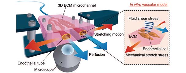
Concept of an ECM-based stretchable microfluidic system. The system can apply fluid shear stress and stretch stress to ECs cultured in 3D ECM environment with real-time fluorescent imaging.
In this approach, the mold was first patterned with bifurcations and cascading dimensions as low as 0.2 mm in width. A commercially and ubiquitously available fused deposition modeling (FDM) 3D printer was used to print the sacrificial mold made with polyvinyl alcohol (PVA). Unlike a well-establish method such as replica molding where multiple steps of assembly and alignment were required to create microchannel with 3D geometry, sacrificial molding enabled the rapid fabrication of microchannels in various matrices. The mold was embedded entirely in an ECM (gelatin), cured with transglutaminase; sealing, alignment, or stacking were not necessary when making the platform for blood vessels and surrounding tissue.
“Since the PVA mold is removable in water, the process of fabrication was entirely completed using only water. This is important to ensure the biocompatibility of the fabricated microchannels,” said Jason Goh, a PhD scholar at SUTD.
“Sacrificial molding of a fused deposition modelling 3D-printed mold offers wide freedom of design and potentiates the fabrication of more physiological relevant platform,” added Assistant Professor Michinao Hashimoto from SUTD.
Human endothelial cells were readily cultured on the surface of the microchannel to form a tube mimicking blood vessels. The hallmark behavior of blood vessels, such as its pulsatile flow, was successfully achieved under conditions of perfusion and stretch. This blood vessel platform served to broaden the spectrum of applicability of current vascular in vitro models to investigate pathological conditions in a more physiologically relevant manner.
“We successfully demonstrated to engineer substitutes for blood vessels with sufficient mechanical strength to withstand the applied fluid pressure and stretching present in the human body. The platform will be useful to understand the mechanisms of vascular diseases,” said Azusa Shimizu, the lead author and an MSc student, and Associate Professor Hiroaki Onoe from Keio U, Japan.
The research work has been published and featured prominently in the inside cover of Lab on a chip, the top journal covering original work related to the miniaturization below the microscale and at the interface between technological advancements and impactful applications. Azusa Shimizu (Keio U) collaborated with Jason Goh (SUTD) and Shun Itai (Keio U). Other senior researchers of the project include Dr Shigenori Miura from the University of Tokyo.
Reference:
ECM-based microchannel for culturing in vitro vascular tissues with simultaneous perfusion and stretch, Lab on a Chip (DOI: 10.1039/d0lc00254b)
Acknowledgements:
This work was partly supported by Grant-in Aid for Scientific Research (B) (19H04440) from Japan Society for the Promotion of Science (JSPS), Japan, and the research grant from Digital Manufacturing and Design Centre (DManD) at Singapore University of Technology and Design (SUTD) (RGDM1620403), Singapore.