SUTD researchers develop liquid metal antenna that can conform to soft biological tissues
SUTD - Kento Yamagishi, Terry Ching, Wenshen Zhou, Shao Ying Huang and Michinao Hashimoto
National University of Singapore - Terry Ching
Direct-ink-writing-based 3D printing of microchannels on a 7 μm-thick elastomeric substrate was demonstrated to fabricate liquid metal microfluidic antennas with unprecedented deformability and tissue-adhesion.
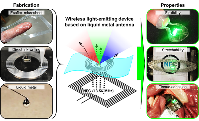 Image: Development of a wirelessly powered light-emitting device based on a liquid metal microfluidic antenna. Direct ink writing (DIW) of silicone microchannels on an Ecoflex microsheet (7 μm thick) was followed by the injection of gallium-based liquid metal — Galinstan. The fabricated device showed unprecedented flexibility, stretchability, and conformal tissue adhesiveness.
Battery-free, wirelessly powered electronic devices have been developed for use in advanced medicine and healthcare. However, such electronic devices interfaced with the biological tissues are required to have an antenna for wireless communication and energy transfer.
To place devices on biological tissues, flexible and stretchable electronics technologies can solve the mechanical mismatch at the tissue-device interface. To mechanically follow and match the curvature of organs, such devices employ a thin-film design composed of elastomers to ensure bendability and stretchability.
To improve the electrical and mechanical performance of antennas for the wireless devices interfaced with tissues, researchers at the Singapore University of Technology and Design (SUTD) used Galinstan, a low-toxicity liquid metal, to create stretchable and conductive traces for the coil.
The developed Galinstan-based microfluidic antenna, published in Advanced Materials, maintains high wireless powering efficiency even when subjected to extreme deformations such as stretching, bending, and twisting.
The device can also conformally adhere to dynamically moving moist and soft biological tissues, serving as a thin wireless platform for implantable devices. This technology "can advance implantable medical applications in hard-to-reach lesions with conventional devices," said Dr Kento Yamagishi, the lead author of the study.
Direct-ink-writing 3D printing of microfluidic antennas
Existing liquid metal antennas have monolithic structures with >100 µm of thickness, but are still too thick to be flexible enough to conformally follow the surface of biological tissues. The research group proposed to use direct ink writing (DIW) 3D printing to create softer, non-monolithic structures to address this problem. A fast-curing silicone sealant was pneumatically extruded onto a 7-µm thick elastomeric substrate (Ecoflex microsheet) to pattern the outline of the microchannel. DIW 3D printing enabled controlling the width, space, and height of the antenna. After embedding the light-emitting diodes (LEDs) and jumper wiring, the outline was sealed with a free-standing Ecoflex microsheet to form microfluidic channels.
A sacrificial layer of polyvinyl alcohol (PVA), a water-soluble polymer, was used to provide mechanical support and enabled the liquid metal to flow (download supplementary video) in the thin-film microchannel to form the stretchable coil. The fluidic antenna (Fig. 1) operates at a frequency close to 13.56 MHz, the standard near field communication (NFC) frequency. The liquid metal antenna offered a high quality (Q)-factor (>20), demonstrating the efficiency of wireless powering.
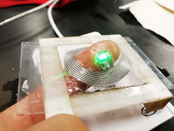 Fig. 1: Image of the free-standing light-emitting thin-film device. Embedded LED chips were wirelessly turned on when the antenna was deformed by a finger.
Flexibility, stretchability, and conformal adhesion to moist and soft biological tissues
Wavy and serpentine patterns of solid metallic circuits are often used in stretchable electronics to ensure the elongation required to yield the electrical connections under strain. However, there is a limit to elongation—the wiring eventually fractures. On the other hand, liquid metal offers unlimited stretchability, making it an attractive choice for experiencing large deformations.
The research group reported that the Galinstan antenna can experience up to 200% tensile strain, match a 3 mm radius of curvature, and withstand a 180o twisting angle while maintaining a high Q factor. Repetitive tensile strain tests (download supplementary video) showed no degradation in the Q factor or meaningful shift in the operating frequency, highlighting the mechanical stability of the device.
Lastly, to enhance the adhesiveness of the Galinstan antenna to the moist and soft biological tissues, polydopamine—a mussel-inspired bioadhesive—was used to enhance the adhesion strength to avoid sutures that causes damages to the tissue. Demonstrations using an explanted porcine small intestine (download supplementary video), heart (download supplementary video) (Fig. 2), and inside a chicken leg (download supplementary video) revealed the stable adhesion and reliable wireless function of the antenna even under deformations of the tissues.
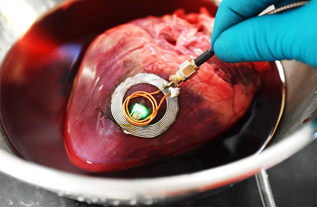 Fig. 2: Image of the light-emitting device attached to the surface of an explanted porcine heart. LEDs were wirelessly turned on by holding the transmission coil (power supply:12 MHz, 1 W) near the antenna.
“Our liquid metal antenna offers a new capability for the design and fabrication of wireless biodevices, which require conformal tissue-device integration. We believe this technology paves the way towards minimally invasive, imperceptible medical treatments,” said Dr Kento Yamagishi, the lead author of the paper.
“While we demonstrated the direct fabrication of microchannels on ultrathin films in this work, direct 3D printing of microchannels enables the creation of microchannels and other fluidic components on different types of surfaces, including biological surfaces. We believe that such capabilities will bring new opportunities for biological sensing, communication, and therapeutics,” added Principal Investigator, Associate Professor Michinao Hashimoto.
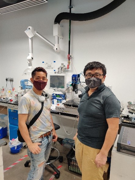 Lead author Dr Kento Yamagishi (left) and corresponding author Associate Professor Michinao Hashimoto (right) from SUTD’s Soft Fluidics Laboratory.
Acknowledgements:
This research project was led by SUTD's Soft Fluidics Laboratory (Facebook: SoftFluidics, Twitter: softfluidics), in collaboration with Associate Professor Shao Ying Huang’s group from SUTD. The research work has been published in Advanced Materials, a peer-reviewed high-impact journal (impact factor: 27.398) covering materials science, published by Wiley-VCH.
Reference:
Ultra-Deformable and Tissue-Adhesive Liquid Metal Antennas with High Wireless Powering Efficiency, Advanced Materials. (DOI: 10.1002/adma.202008062)